The Buzz on Babyecho
The Buzz on Babyecho
Blog Article
Get This Report on Babyecho
Table of Contents3 Easy Facts About Babyecho DescribedBabyecho - An OverviewThe Definitive Guide for BabyechoAll About BabyechoNot known Details About Babyecho The Ultimate Guide To BabyechoAll about Babyecho

A c-section is surgical procedure in which your infant is born through a cut that your medical professional makes in your stomach and uterus. No issue what an ultrasound reveals, talk with your copyright regarding the best look after you and your infant - fetal doppler at home. Last examined: October, 2019
During this check, they will certainly check the infant is expanding in the right location, whether there is greater than one infant and they will also inspect your child's development up until now. This screening is offered between 10 14 weeks of pregnancy and is utilized to assess the opportunities of your infant being birthed with one or more of these conditions.
Not known Details About Babyecho
It entails a mixed examination of an ultrasound check and a blood examination. Throughout the check, the sonographer will gauge the liquid at the rear of the baby's neck to determine 'nuchal translucency' - https://dzone.com/users/5144371/babydoppler1.html. They will then determine the opportunity of your infant having Down's, Edwards' or Patau's syndrome using your age, the blood test and scan results
Throughout this check, the sonographer look for architectural and developing problems in the infant. During this scan visit, you may be offered testings for HIV, syphilis and hepatitis B by a professional midwife. Sometimes, a third-trimester check is advised by your midwife adhering to the outcomes of previous tests, previous complications or existing medical conditions.
The typical 2D ultrasound generates level and described photos which can be utilized to see your infant's internal body organs and aid detect any kind of interior problems. These black and white images aid the sonographer figure out the infant's gestation, development, heart beat, development and dimension. Some pregnant mothers pick to have a 3D ultrasound scan due to the fact that they reveal more of a real-life picture of the infant.
The Of Babyecho
3D ultrasound scans show still images of your baby's exterior body instead than their withins, so you can see the shape of the infant's face functions. 4D ultrasound scans are comparable to 3D scans yet they reveal a moving video clip rather than still pictures. This records highlights and darkness much better, as a result developing a clearer picture of the baby's face and motions.

or (8-11 weeks) (11-14 weeks) (14-18 weeks) (19-23 weeks) or (24-42 weeks) Recommended at Optional -, extra regularly in some problems This this article scan is done to and to establish an (EDD). A is identified throughout this check. The majority of moms and dads select this check for. Likewise is necessary before the blood examination called as (NIPT) to determine the.
How Babyecho can Save You Time, Stress, and Money.
Occasionally a might be required to get and a clearer image. This is usually done and periodically a might be required (baby heartbeat doppler). Verify that the baby's heart is present; To a lot more accurately.
Please see below. These scans might be done, nonetheless some of the and thus, a is required to This check is done generally at.
How Babyecho can Save You Time, Stress, and Money.

Additionally, the can be by by an. () The method nearer the is valuable to. Occasionally, an which was previously may be.
The 4-Minute Rule for Babyecho
If, these scans may be to. on the findings, a may be used. During all the, a 3D check (of the child) can additionally be executed. The is dependent on the position of the,,, amount of and. This includes, along with; This consists of, together with (14-20 weeks).
A scan is vital prior to this test is done. If you're looking for, prepare an assessment with Dr Sankaran through her. Obstetrics & gynaecology in London.
More About Babyecho
The examination can supply valuable details, assisting females and their health-care carriers handle and care for the maternity and the fetus.
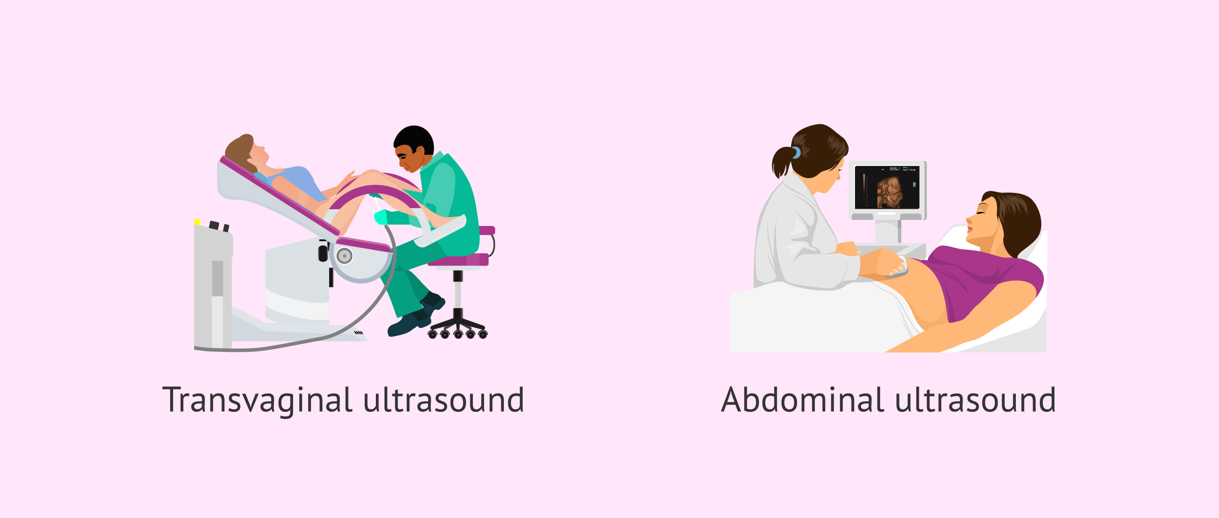
A transvaginal ultrasound creates a sharper photo and is often used in very early pregnancy. Ultrasound devices are regarding the size of a grocery store cart.
Report this page